Instructions
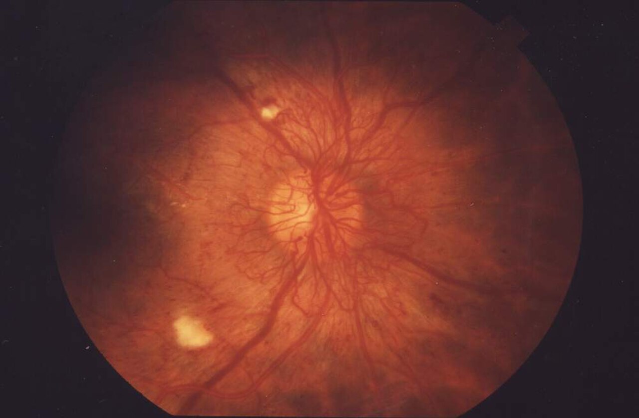
You are a medical student on placement in the diabetes clinic. Yiannis Georgiou, 61, who has type 2 diabetes, is here for the results of his eye photographs. They are below.
Interpret the fundus photographs
Explain the results and your management plan to the patient
Right macula (report)
Ring of hard exudates within one disc diameter of the fovea; dull foveal reflex
Medicines
Metformin 1 g twice daily, gliclazide MR 60 mg, amlodipine 5 mg; smokes 10 a day
Fundus photographs (non-mydriatic retinal camera) and visual acuity
| Item | Value | Reference range |
|---|---|---|
| Visual acuity, right eye | 6/12, no improvement with pinhole | 6/6 |
| Visual acuity, left eye | 6/9, improving to 6/6 with pinhole | 6/6 |
| Left eye: red reflex | Clear | Clear |
| Left eye: disc | Normal colour, margins and cup; no abnormal vessels | Normal |
| Left eye: vessels | Normal calibre; no beading | Normal |
| Left eye: macula | Normal foveal reflex; no deposits within two disc diameters of the fovea | Normal |
| Left eye: periphery | About a dozen tiny red dots and small dot haemorrhages, mostly nasal to the disc; two small clusters of waxy yellow deposits in the far periphery; no fluffy white patches | None |
Recent bloods (GP, 2 weeks ago): HbA1c 9.5% (80 mmol/mol); eGFR 62 mL/min/1.73m2; urine albumin-creatinine ratio 8 mg/mmol; total cholesterol 5.6, LDL 3.4, HDL 1.0, triglycerides 2.8 mmol/L.